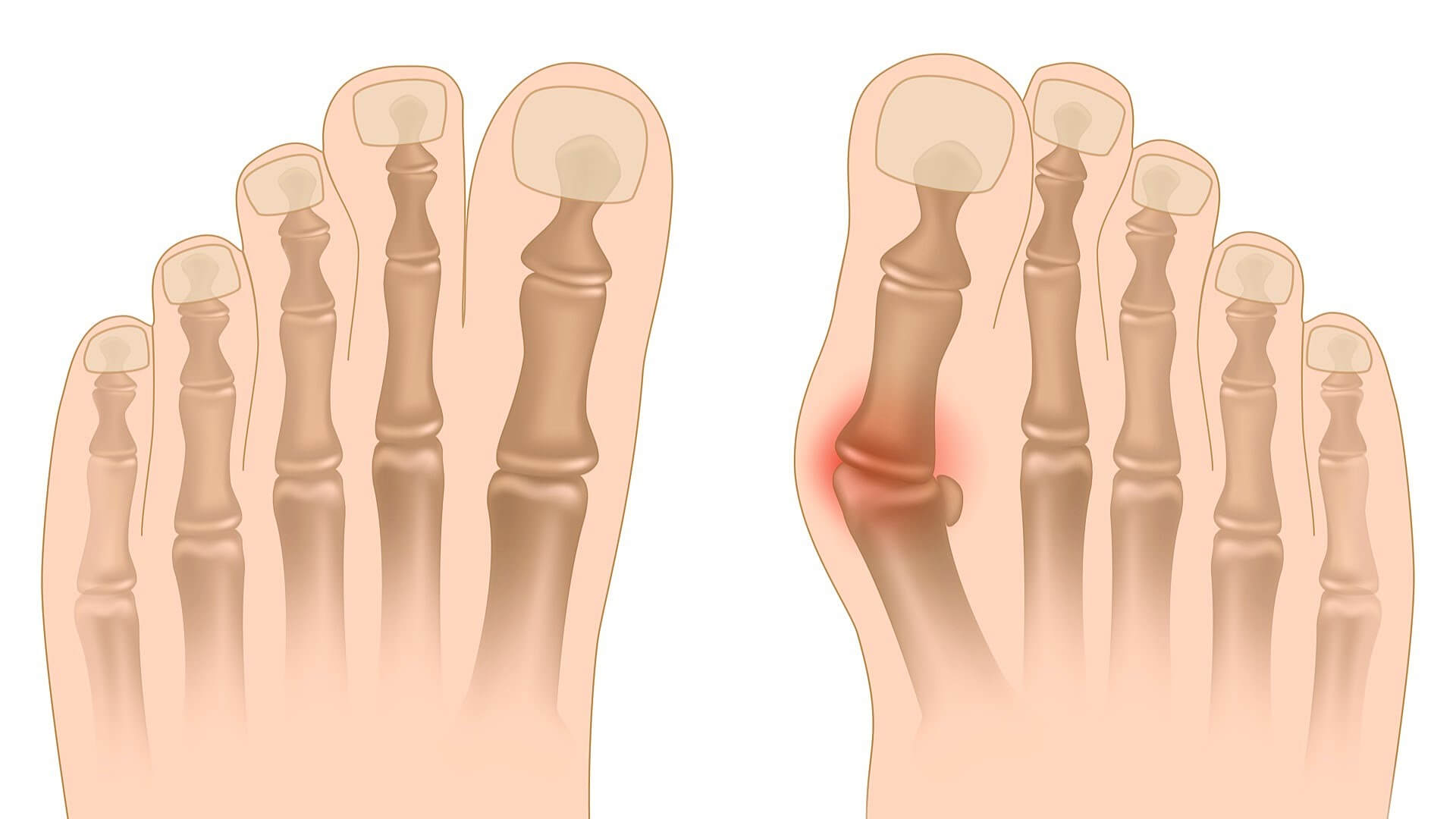
Quick answer: If you have a bunion, start with the basics: wear roomy, low-heeled shoes with a wide toe box, use padding or a night splint to ease pressure, apply ice after activity, and choose pain relief you can use safely. These steps relieve pain and slow progression — they do not remove the bump. If pain persists or walking becomes difficult, see a podiatrist to discuss imaging and whether surgery is appropriate. This page explains what a bunion is, its anatomy and causes, and the full range of conservative care.
⚠️ When to see a doctor: If you have diabetes or poor circulation, reduced feeling in your feet, signs of infection (redness, warmth, swelling, an open wound or discharge), or severe or worsening pain, seek medical care promptly rather than relying on home treatment.
Well, many people all around the world are complaining about having a bunion and of all the people, women are comparatively higher in proportion.
Before getting into further details of how and why the bunions are formed and what to do once it develops, first of all, we need to understand what exactly bunion is?
So, letâ··s start.
What is a bunion?
A bunion is a deformity of the foot that could be caused due to a number of reasons. The term bunion is basically a layman term used for â··HALLUX VALGUSâ··. Hallux Valgus is a Latin term, â··the big toeâ·· and valgus means that â··turned
away from the midlineâ··.
So, a bunion can be defined as:
â··Deformity of the foot in which the big toe is turned away from its normal midline orientation.â··
Although it sounds like just having a bump on the foot but in reality if we look into it, there is a progressive change in the normal bony alignment of the foot, which can also be seen over the internet on Wikipedia, describing bunions as â··the deviation of the big toe towards the midline of the 2nd toe accompanied by the deviation of the first metatarsal bone towards the normal midlineâ··â··.
Bunions are not formed just within a day or two, it grows gradually over the time and it takes much longer to be prominent enough to bring visible differences in the actual alignment of the foot.
There is another condition called the Tailorâ··s bunion, which is often misdiagnosed with actual bunions (Hallux Valgus). It differs from Hallux Valgus in terms of its location, being present at the base of little toe on the outside of the foot. So, to understand better that how a bunion is formed and what causes it to occur, we need to take a look at the anatomy of the bunion.
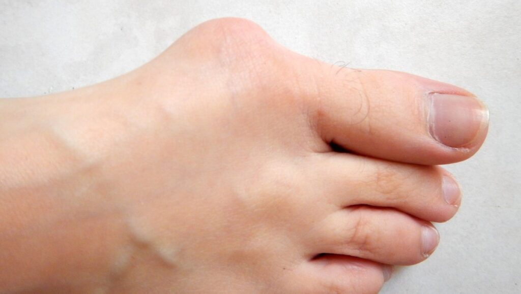
Anatomy Of Bunion
A bunion usually develops when there is a stressing force applied repeatedly on the foot causing the foot to squeeze together. As a result, the big toe of the foot is pushed against the other toes and end up displacing either over it or under it and the MTP joint angles out from the foot leading to its misalignment.
Although the major joint involved in Hallux Valgus(HV) is 1st MTP joint, but while making a diagnosis some other areas need to be examined too as they also get involved with time and severity of the deformity and become an important part of considerations of management and treatment plans like planning a surgery or Physical therapy sessions.
The other associated areas are:
- Interphalangeal joints (IJP)
- 1st metatarsocuneiform joint (MTCJ)
- Proximal Phalanx Articular Angle (PPAA)
- Distal Metatarsal Articular Angle(DMAA)
However, the deformity in all the associated areas doesnâ··t occur simultaneously. It develops gradually and involves other structures in a step-like manner.
First of all, the failure of medial structures surrounding the 1st MTPJ occurs. The medial structures involve:
- Medial collateral ligament
- Medial sesamoid
When the medial structures fail, the causes a number of events to take place leading to the involvement of multiple joints and structure.
The sequel of the events are;
- MT head moves medially, crossing over the sesamoid apparatus.
- The medial sesamoid, in turn, erodes the crista and the plantar facing cartilage.
- The proximal phalanx falls into valgus position.
- The pressure on the bursa over the medial aspect of joint increases leading to the thickening of the bursa.
- The tendon of the certain muscles attains a new position thus worsening the condition.
- Flexor and Extensor hallucis tendons bowstring across the joint.
- Abductor hallucis tendon also changes its position with the pronation of Toe and its force further adds to the pronation.
Cause Of Bunion
There could be a number reasons which can be the culprit in the formation of bunions and multiple factors that can aggravate the existing problem.
Some key factors to be considered are as following:
Hereditary cause: Bunion has a genetic predisposition that means that it can run in the families. Many people can have congenital faults in the foot shape leading to the formation of a bunion. Most commonly occurring congenital foot problems are:
Pes planus: That refers to the Flat feet, meaning the normal foot arch is absent.
Low arches of the foot.
Loose joints and tendons.
Defects in the anatomy of the first toe: If the head of the 1st metatarsal bone is too round it decreases the stability of the joint and makes it more vulnerable to be deformed when increased external forces are applied.
Hypermobility of the joint.
Mal footwears: The second most commonly occurring cause of the bunion is poor and cheap footwear. Shoes which are too tight and have narrow and pointed toes result in additional pressure on the toes. The increased pressure for the longer period of time causes the disturbance in the normal alignment of the foot.
Similarly, high heels are also found to exacerbate the bunion formation because it causes the body weight to shift to the forefoot pushing the toes in the front of the shoes. This is the main reason why the incidence of Hallux Valgus is greater in women than men.
Inflammatory conditions: Certain degenerative changes that lead to the inflammatory conditions like arthritis can be responsible for the bunion formation of an individual. Treating inflammation relieves symptoms, but it does not reverse arthritic changes or the bunion itself.
Occupation-related stresses: The people who are on the field for longer durationâ··s of time and those who are involved in professions like athletics, sales workers, teachers which demand a lot of standing or walking or increased impact forces are more prone to form bunions.
Pregnancy: Hormonal changes during pregnancy are responsible for the bunion formation and aggravation of the condition. It is associated with loosening of the ligaments and tendons due to hormonal changes causing the foot to lose its normal arch and become relatively flat.
Obesity: Obesity although doesnâ··t cause the bunion formation itself but it is found to play some part in triggering the already present condition.
Age: The age plays an important role too. The peak age for the bunion to occur is 30-60 years. However, it can affect the young girls of 10-15 years, this condition is called Adolescent bunion.
Symptoms Of Bunion
Symptoms associated with Hallux Valgus do not usually occur in the early stages. But with the progression of deformity, as more structure starts to get involved and condition starts to worsen, the symptoms become more prominent and visible. The patients usually present with:
- Severe Pain in the toes.
- Erythema (Redness) of the affected area.
- Swelling Of 1st MTP joint.
- Numbness of the toe.
- Tenderness around the toe.
- Burning sensation.
- Difficulty standing and walking (in advanced stage)
- Associated systemic diseases.
- Abnormal Gait.
On Examination, following abnormalities are found to be present.
- Lateral deviation of the 1st MTP joint
- Weakness of hallux abductor muscles·Hypermobility of the first metatarsocunieform joint
- Chronic Achilles tendon tightness
- Toe pronation deformity
- Failure to supinate the foot
- Lack of weight bearing on the hindfoot
Radiological Findings
The final diagnosis and treatment considerations are formed on the basis of radiological findings. Best radiographs are:
- X-ray of foot in standing position (in the Anteroposterior (AP) and Lateral planes)
- X-ray of foot in non-standing position (oblique view)
- Sesamoid view (optional)
Distal Metatarsal Articular Angle(DMAA): It shows the orientation of the articular surface of the head of MT in relation to the 1st MTPP.
Normal value: <10 degrees
Hallux valgus: increased
Proximal Phalanx Articular Angle (PPAA): angle formed by the intersection of a line along the long axis of the PP and a line along the proximal joint surface of the PP
Hallux Valgus Angle(HVA): The angle formed by the intersection of the lines along the long axis of the PP and the 1st· MT.
Normal:<15 degrees
Intermetatarsal Angles(IMA): The angle formed by the intersection of long axis lines along the first and 2nd· MT.
Normal:<9 degrees.
Impairments Caused By Bunion
The MTP joint is the main weight-bearing joint of the foot. When a bunion is formed in this area, it affects all the functional activities of the foot as multiple structures get involved, thus producing a major hindrance in the ADLs and IADLs.
Following the pattern of impairments occur commonly:
- Bunion affects the big toe which in turn puts pressure on the other toes.
- Severe pain causes the avoidance to shift weight on the big toe.
- There is increased pressure on the other toe thus the toes get misaligned and end up developing hammertoes.
- Resultantly, the standing, walking and other activities of daily living get affected.
- The overall quality of life declines, which leads to the anxiety, depression and mental irritation
So, it Is very important to pay attention to the bunion and get it corrected as soon as possible.
Treatments
There are two types of interventions for the bunion i.e surgical and conservative. In the initial stages, there is no need for the surgical interventions. The treatment is directed to relieve the pain and prevent the further deterioration of the joint and progression of the deformity.
The best results can be obtained with the conservative management until and unless the condition becomes so severe that pain becomes debilitating and doesnâ··t improve with non-surgical management and there are multiple joint deformities. In this case, surgical intervention is the only solution.
Conservative management of bunion includes:
Lifestyle modifications: The factors that are contributing to the development and exacerbation of the condition need to be controlled. Such as
- Controlling obesity
- Wearing ergonomically suitable shoes: that are neither too tight nor too narrow to cause excessive pressure forces on the toes.
- High heels should be avoided as much as possible and should be worn only occasionally to protect the feet from damaging forces.
- Occupation related stresses should be minimized by taking rest breaks more often during working hours and avoiding constant long standing.
Avoiding pain aggravating activities: such as prolonged standing, walking, running, cycling, thus all those activities that may put excessive stress on the already damaged toes.
Cryotherapy: It is applied for pain relief and reduction in inflammation. Cryotherapy means applying ice to the affected area. It numbs the joint and can ease pain and inflammation after activity — about 15–20 minutes at a time, with a cloth between the ice and your skin.
Bunion Pads: ·It is used to prevent Progression of Deformity and avoid pain. Bunion pads are specially designed pads usually filled with some gelatinous material which help to decrease the impact forces on the foot when it strikes the ground and ensures the correct positioning of the foot during standing and walking.
Analgesics: Acetaminophen and other NSAIDs like ibuprofen and aspirin can be used to reduce inflammation and pain.
Corticosteroids injection(cortisone): It also helps provide pain relief. But due to its other side effects, it is not often used until in the case of a severe
Orthoses: Custom made orthoses are used to prevent exacerbation of condition. The orthoses are fitted in the shoe to keep the foot in the normal alignment. But orthoses seem to help only in the only stages of the deformity (roughly up to 6 months) after that no difference in the control of the disease is observed.
Physiotherapy:
It is the mainstay of treatment, it includes:
Extension Exercise Of The Toes:
Any simple task involving extension of the toe can be suggested to the patient to practice extension exercises e.g drop a piece of cloth or any small object on the floor and ask the patient to pick the object with the affected toe and lift it up as high as he can without lifting the heel from the ground.
- Grade 3 joint mobilizations (Abduction and flexion)
- Muscle and joint stretching.
- Bunion stretch and soft tissue release
- Ankle dorsiflexion exercises.
- Strengthening of leg muscles
Toe Towel Curls:
Put a towel down on the floor and stand straight on it. Then try to curl the toes in a way that you pull the towel toward you with your toes. Do 10 repetitions and then try to scoot the towel away from you with the help of toes again. It strengthens the intrinsic muscles of the foot that holds the arch of the foot.
Taping:
KT tape is a specially designed tape, it corrects the misaligned MTP joint and thus helps relieve pain and pressure over the toes.
Postural training program for the foot.
Gait training.
Electrotherapy: Modalities including Ultrasound, Short-wave diathermy may provide some relief to the pain.
whirlpool therapy.
Massage.
Educational only — not medical advice. This article was originally published on Footcarely in 2018 and has been reviewed and updated against current guidance. See our Medical Disclaimer and How We Review pages.